What Are the Methods to Biopsy a Lump in the Lung?
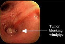
Bronchoscopy. A lighted tube that the physician can see through is placed into the windpipe of a patient who has had sedation and/or anesthesia. The physician can identify visually the region to perform a biopsy. This technique also provides information whether the tumor may be removable by surgery. It works best for tumors near the central windpipes. Usually a same day procedure.
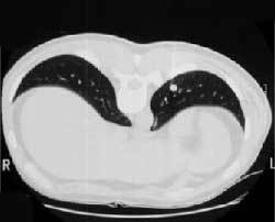
CT Scan Biopsy. The patient is placed into the CT scan machine and using X-ray guidance, the area in question is biopsied using a thin needle placed through skin that has been carefully anesthetized. It works best for tumors near the edge of the lung. Same day procedure.
Mediastinoscopy or mediastinotomy. When a node near the air passages must be biopsied, a mediastinoscopy may be used to make exact biopsies of the glands around the airway. Some glands in the left chest cannot be reached by the mediastinoscopy introduced into a a low neck incision; a mediastinotomy (Chamberlain procedure) can be used if needed to reach a suspicious gland in the left chest. A small incision is made near the breastbone and a small piece of rib is removed to allow access to that area.
Fluid cytology. If the patient is coughing up abnormal phlegm, has abnormal amounts of fluid around the lung, or has an enlarged gland elsewhere, then a sputum sample or a needle placed in these other abnormal areas can obtain tumor cells to make a diagnosis.
Operation. Sometimes, less invasive methods to confirm the cause of the lung lump fail and surgery is required. A definite diagnosis is not always needed before an operation, such as when the risk of the lump being cancer is high, the risk of an operation is low, and an operation to potentially cure the patient would be appropriate. The most efficient means to obtain a diagnosis is a biopsy at the same time that the definitive operation is carried out.
Thoracoscopy. In this method, the lump in the lung is removed or biopsied with a special needle. See Treatment. If no more surgery than a biopsy is needed, many patients can leave the hospital the next day.
EUS (endoscopic ultrasound) or EBUS (endobronchial ultrasound) Specially designed ultrasound probes attached to small tubes that are placed in the swallowing tube or windpipe allow biopsy of certain deep lymph nodes that formerly would require a surgical incision to access.
Navigational Bronchoscopy (including Robotic) The patient has a special CT scan to create a three-dimensional "map" of their lungs and chest. Then, using a miniature version of GPS technology that is used to navigate the world, a special probe can precisely find the target within the patient's chest.